Intravascular imaging is a critical tool in modern cardiology, enabling accurate diagnosis and therapeutic guidance in patients with coronary artery disease.
The three main methods of intravascular imaging are Intravascular Ultrasound (IVUS), Optical Coherence Tomography (OCT), and Near-Infrared Spectroscopy (NIRS). Each of these techniques offers unique advantages for the assessment of coronary vessels.
What is intravascular imaging?
Intravascular imaging refers to techniques that utilize catheters to visualize the interior walls of the coronary arteries. These methods allow for a detailed assessment of the structure and composition of the vessel wall, qualitative and quantitative analysis of atherosclerotic plaques, and evaluation of associated luminal stenoses.
Intravascular imaging is extremely important for diagnosing underlying coronary artery disease and for guiding therapeutic interventions, such as stent placement, ultimately improving the outcomes of such treatments.
What is Optical Coherence Tomography (OCT)?
Optical Coherence Tomography (OCT) is an advanced imaging technique that uses light emission through a fiber-optic cable to create high-resolution images of the vessel’s inner wall. It is based on analyzing light reflections from various vascular wall structures using a rotating optical fiber.
OCT provides images with a resolution comparable to that of light microscopy, allowing for precise evaluation of plaque quality and other microscopic vascular structures. This technique is particularly useful in accurately assessing atherosclerotic plaques and optimizing stent deployment.
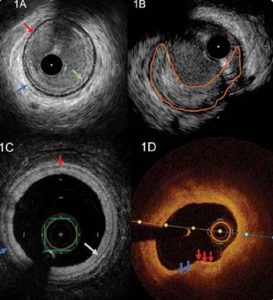
- (1A) Normal vessel imaging with IVUS: The three layers are visible—intima (green arrow), media/EEL (black echolucent ring – red arrow), and adventitia (blue arrow).
- (1B) Eccentric atherosclerotic plaque visualized by IVUS (orange-framed area): IVUS’s depth penetration allows for accurate assessment of plaque thickness.
- (1C) Normal vessel imaging with OCT: The three layers are visible—intima (green arrow), media/EEL (black echolucent ring – red arrow), and adventitia (blue arrow).
- (1D) Eccentric atherosclerotic plaque in OCT (arrow-marked area): OCT’s limited penetration does not allow accurate plaque thickness assessment (due to light absorption). However, its high resolution enables identification of features like macrophages (granular, high backscatter structures – red arrows).
What is Near-Infrared Spectroscopy (NIRS)?
Near-Infrared Spectroscopy (NIRS) enables the identification and quantification of the lipid content of atherosclerotic plaques by analyzing their lipid-rich core. The technique uses a catheter equipped with a fiber-optic component that emits near-infrared wavelength light to analyze the plaque’s chemical composition, providing information on the size of the lipid core within the plaque.
This information is clinically important, as larger lipid cores and higher lipid content indicate more vulnerable plaques, increasing the risk of rupture and consequently acute myocardial infarction.
NIRS is the only imaging modality approved by the U.S. Food and Drug Administration (FDA) for identifying high-risk/vulnerable atherosclerotic plaques. Recent clinical studies have shown that preventive treatment of these plaques with stent placement can significantly reduce the risk of acute myocardial infarction.
What is Intravascular Ultrasound (IVUS)?
Intravascular Ultrasound (IVUS) is a technique that uses ultrasound waves to image the interior of coronary vessels. A catheter with an ultrasound probe is inserted into the vessel lumen. The probe emits ultrasound waves, and the reflected and absorbed signals from the different vessel wall layers create high-resolution cross-sectional images of the coronary arteries, allowing for diagnosis and identification of atherosclerotic plaques and associated luminal stenoses.
IVUS allows precise measurement of the vessel dimensions, composition of the wall and plaque (calcified or soft), total plaque burden, and minimum luminal area (MLA). It is especially valuable for guiding percutaneous coronary interventions (PCI) and stent implantation, helping optimize treatment outcomes.
Where are the examinations performed?
IVUS, OCT, and NIRS examinations are conducted at specialized cardiology centers and hospitals equipped with the necessary technology and experienced personnel trained in both the execution and interpretation of these tests.
Are there any complications from these tests?
IVUS, OCT, and NIRS are generally considered safe procedures, with the risk of serious complications being extremely low (<0.1%) when performed by experienced interventional cardiologists.
What is the cost of the examinations?
The cost of IVUS, OCT, and NIRS may vary depending on the type of equipment (catheters) used, the specific test performed, and the services provided during the procedure.
Overall, IVUS, OCT, and NIRS are invaluable tools for the accurate diagnosis and management of coronary artery disease, offering critical information for targeted treatment and prevention of cardiovascular events.